 Form and structure
Reproductive structures
Both sexual and vegetative reproduction can be found amongst the lichens and, when you look at a lichen with a naked eye or hand lens, you are likely to see some of the structures that act to produce or disperse sexual or vegetative propagules. The aim of this page is simply to describe such structures rather than to deal with the mechanics of propagule production or dispersal but you can find more information about that aspect on the REPRODUCTION & DISPERSAL page. Much of this page will be devoted to the structures involved in sexual reproduction. When it comes to vegetative reproduction the relevant structures are far less obvious. Indeed, sometimes you may wonder whether you're looking at a definite structure or just a modification of thallus morphology and some types of vegetative propagules are released with the development of no structure to help produce and disperse such propagules.
Finally, in almost all lichens the mycobionts are ascomycetes and this page will be devoted to the reproductive structures of the ascolichens. There is a separate page about BASIDIOLICHENS which contains details of both their vegetative and reproductive structures. Much of the information on this page has been taken from the sources given in the following reference button![]() .
.
Sexual reproduction
Only the fungal partner reproduces sexually.
The most commonly seen sexual reproduction structures are apothecia. These are typically circular and disc-like or cup-like though there are also species in which the apothecial surface bulges outward. They may be of the same colour as the thallus ![]() or strikingly different
or strikingly different ![]() and vary in diameter from under a millimetre to over two centimetres, depending on species, with the larger apothecia found on the more substantial foliose lichens. In most species the apothecia sit on the thallus surface but there are species in which the apothecia are immersed in the thallus
and vary in diameter from under a millimetre to over two centimetres, depending on species, with the larger apothecia found on the more substantial foliose lichens. In most species the apothecia sit on the thallus surface but there are species in which the apothecia are immersed in the thallus ![]() and others where the apothecia are raised on stems, from a millimetre or two
and others where the apothecia are raised on stems, from a millimetre or two ![]() to over a centimetre in length
to over a centimetre in length ![]() . The crustose and foliose lichens have distinct lower (or ventral) and upper (or dorsal) surfaces, the former facing towards the substrate the latter away. Apothecia are almost always found on the upper thallus surfaces but the foliose genus Nephroma
. The crustose and foliose lichens have distinct lower (or ventral) and upper (or dorsal) surfaces, the former facing towards the substrate the latter away. Apothecia are almost always found on the upper thallus surfaces but the foliose genus Nephroma ![]() is an exception. In this photo
is an exception. In this photo ![]() of a species of Peltigera the apothecia are the dark brown areas on the upright parts of the thallus.
of a species of Peltigera the apothecia are the dark brown areas on the upright parts of the thallus.
The following diagram shows a cross-section of an apothecium. Apothecia vary in internal structure but this diagram shows some basic features. The hymenium is a layer composed of asci interspersed with sterile hyphae (called paraphyses). There are five asci to be seen. The rightmost two I have left untouched. I have used red to highlight the wall of the middle ascus and I have used red to highlight spores in the remaining two asci. The rest of the apothecium serves to support the hymenium and you can see that there can be differences in the tissues within an apothecium. The hymenial surface is referred to as the disc of the apothecium. In many lichens the apothecial margins are composed solely of fungal tissue (typically different from the paraphyses and asci of the hymenium) though there are also many species in which the apothecial margin bears an extra layer, composed of fungal and photobiont cells, derived from the thallus and here we refer to a thalline margin. In a species where the apothecia have thalline margins the apothecial margin is of a different colour to that of the disc, as you can see in the apothecia of Haematomma africanum ![]() and Tephromela atra
and Tephromela atra ![]()
![]() .
.

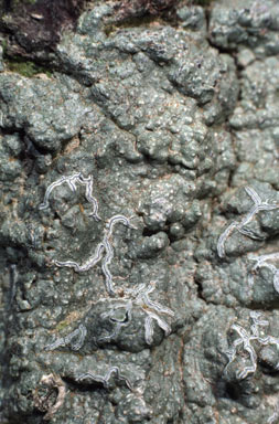
While the majority of apothecia are persistent and may still be present long after the spores have been dispersed, the apothecial tissue in some breaks down to leave a mazaedium, namely a powdery mass of spores and hyphal fragments. This is particularly the case with the lichens of the family Caliciaceae. Another way in which some lichen genera differ from the norm is by having apothecia that are narrow and elongated ![]()
![]() (sometimes even branching ). These are termed lirellae and are found in the genera of the family Graphidaceae.
(sometimes even branching ). These are termed lirellae and are found in the genera of the family Graphidaceae.
Another type of fungal fruiting body is a perithecium which, to the naked eye, looks like a small, hemispherical pimple, typically black ![]() and varying in diameter from under a millimetre to about two millimetres. Depending on the species perithecia may develop totally on the lichen thallus or embedded in the thallus. Perithecia often develop separately but in some genera a number of perithecia will develop within a stroma, namely a matrix of fungal hyphae, often pigmented black. Within a perithecium there is a typically spherical chamber, with a hole (or ostiole) in the roof of the chamber and asci and paraphyses on the floor of the chamber.
and varying in diameter from under a millimetre to about two millimetres. Depending on the species perithecia may develop totally on the lichen thallus or embedded in the thallus. Perithecia often develop separately but in some genera a number of perithecia will develop within a stroma, namely a matrix of fungal hyphae, often pigmented black. Within a perithecium there is a typically spherical chamber, with a hole (or ostiole) in the roof of the chamber and asci and paraphyses on the floor of the chamber.
You'll find more diagrams of microscopic features via the SCHNEIDER'S BOOK page.
Vegetative reproduction
The most significant vegetative propagules are isidia and soredia. While they are present on many lichens there are also many species lacking them. Moreover, some species may have both isidia and soredia while others will produce only one type of propagule.
Isidia are small outgrowths of the thallus, from about 50 micrometres to a millimetre or so in length. They contain both fungal hyphae and photobiont cells and vary in shape, depending on species, from bulbous to cylindrical or branched, sometimes even coralloid ![]() . The following diagram illustrates this, showing different types of isidia protruding from the general level of the lichen thallus. The green dots indicate photobiont cells and the grey areas the fungal hyphae. Isidia are often present in large numbers and, at first glance, an isidium-rich part of a thallus can look somewhat indistinct or fuzzy, compared to an isidium-free area. Here
. The following diagram illustrates this, showing different types of isidia protruding from the general level of the lichen thallus. The green dots indicate photobiont cells and the grey areas the fungal hyphae. Isidia are often present in large numbers and, at first glance, an isidium-rich part of a thallus can look somewhat indistinct or fuzzy, compared to an isidium-free area. Here ![]() is a photo of the foliose species Xanthoparmelia ewersii, which bears abundant isidia except near the thallus margins, which are light coloured whereas the dark areas are dense with isidia. Here
is a photo of the foliose species Xanthoparmelia ewersii, which bears abundant isidia except near the thallus margins, which are light coloured whereas the dark areas are dense with isidia. Here ![]() is a closer view of a marginal area where you can see a few sparser isidia and here
is a closer view of a marginal area where you can see a few sparser isidia and here ![]() is a closer view of dense isidia. Broken off isidia may be transported by wind, water or animals (for example, caught on bird feet or in animal fur) and if deposited into a suitable habitat may generate new thalli. In addition to serving as propagules isidia, while still attached to a thallus, effectively increase the surface are of the thallus and so contribute to gas exchange and photosynthesis.
is a closer view of dense isidia. Broken off isidia may be transported by wind, water or animals (for example, caught on bird feet or in animal fur) and if deposited into a suitable habitat may generate new thalli. In addition to serving as propagules isidia, while still attached to a thallus, effectively increase the surface are of the thallus and so contribute to gas exchange and photosynthesis.

Soredia look like small powdery, granules, between about 20 and 100 micrometres in diameter, and each soredium consists of a few photobiont cells surrounded by fungal hyphae. They often originate in the photobiont layer and emerge through cracks or pores in the thallus surface but they also may be produced by a breakdown of the thallus surface. In some species they may appear anywhere on the thallus whereas in others soredium production is confined to delimited areas called soralia and these may take various forms. For example, a soralium may develop as a crack in the thallus surface and so have a linear form or may initially develop as a swelling under the thallus surface but finally break through to finish as a pustulate soralium. Labriform (or lip-like) soralia develop along apices or margins of thallus lobes. A soralium loaded with soredia resembles a tiny, powdery heap. This photo of Pertusaria subventosa ![]() shows many whitish mounds, namely soralia with soredia, on a grey-brown thallus.
The foliose species Parmotrema perlatum
shows many whitish mounds, namely soralia with soredia, on a grey-brown thallus.
The foliose species Parmotrema perlatum ![]() bears soralia near lobe margins.
bears soralia near lobe margins.
As well as propagules in which both the fungal and the photobiont partners are present, it is possible to have vegetative propagules where only one partner is present - but, again, not always in well-defined structures. However a number of lichens do produce asexual fungal propagules in well-defined structures called pycnidia. Superficially a pycnidium resembles a tiny perithecium, in that both are often globose chambers in which propagules are produced. However it is important to remember that the asci in a perithecium produce sexual spores, in which genetic material from two parents is combined, whereas there are neither asci nor genetic combination in pycnidia. To the naked eye pycnidia often look like black pinpricks in the thallus surface and pycnidia may be produced separately or in clusters. That's not to say that every black pinprick indicates a pycnidium since, for example, some black pinpricks would indicate the presence of parasitic fungi. To confirm the presence of lichen pycnidia would require a microscopic examination.
Form and structure pages on this website Crustose lichens |
![An Australian Government Initiative [logo]](/images/austgovt_brown_90px.gif)


