![An Australian Government Initiative [logo]](/images/austgovt_brown_90px.gif)

Types of fungal fruiting bodies (or sporocarps)
Stereoid & paint (skin) fungi
Stereoid fungi (NOT fungi on steroids) often resemble some of the bracket-like polypore fungi, in that there's a thin plate growing out of some wood. Stereum ostrea is a widespread example of this group. In fact, at one time, all members of this group were placed in the genus Stereum - hence the term stereoid.
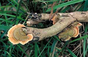 Stereum ostrea |
However, the polypores have pores on the underside but the stereoid fungi have smooth, rather than pored, undersides. Remember that the pores in some of the polypores are very small (10 or more per millimetre). So to tell if you have a polypore or a stereoid fungus, you'll sometimes need to use a hand lens or magnifying glass to check the underside for any pores. Many stereoid fungi start out looking like the paint fungi before they produce the bracket-like growth.
A few stereoid fungi are centrally stemmed, rather then being bracket-like.
Cymatoderma elegans ![]() is an example.
is an example.
Skin or paint fungi.
All of the above examples are markedly three-dimensional. The skin or paint fungi are basically two-dimensional and in many cases one of these fruiting bodies looks just like a layer of paint on the underside of wood lying on the ground. The "paint" layer may be thin or thick, it may be smooth or with some wrinkles, bumps, or very short spines. The technical name for these is corticioid fungi. Here's a species of Exidiopsis, looking like no more than a blue-grey smear on some rotten wood. Hyphodontia australis is another corticioid species, this time with short teeth.
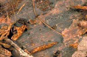 Exidiopsis sp. |
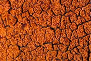 Hyphodontia australis |
Below are links to PDFs of three illustrations of corticioid fungi taken from Scottish Cryptogamic Flora, by Robert Kaye Greville and published in instalments from 1822 to 1826. In each case I'll give first the species name used by Greville and, in brackets, the current name for that species.
Phlebia merismoides (Phlebia radiata)
Figures 1 to 3 show surface views, 4 and 5 show cross sections and spores and asci are in Figure 6.Radulum orbiculare (Basidioradulum radula)
Figures 1 to 4 show whole or parts of fruiting bodies at different magnifications
and spores are shown in Figure 5.Thelephora quercina (Peniophora quercina)
Figure 1 shows several fruiting bodies, the largest with loose margins which lets you see
part of the blackish inner surface (the subject of Figure 2). Spores and asci are shown in Figure 3.
At this time it was believed that all macrofungi produced their spores via asci. In fact the fungi shown here are basidiomycetes yet two of the illustrations show spores in asci, an excellent example of reality not being allowed to get in the way of belief!
The illustrations were printed in black-and-white and then coloured by hand. These particular copies were not for sale but were used as examples for the colourists to follow. At the bottom of the Phlebia merismoides plate you can see a hand-written admonishment: "Color carefully", with no u in the first word.