![An Australian Government Initiative [logo]](/images/austgovt_brown_90px.gif)

|
|
The gametophytes of mosses and leafy liverworts consist of leaves on stems. When you're trying to decide if a leafy bryophyte is a moss or a liverwort, and there are no sporophytes present, the first thing to do is to see if there is a nerve, or costa, in the leaf. If present, a nerve typically shows as a distinct line along the leaf's central axis. The accompanying photo (below, left) (which is an enlargement of part of this photo ![]() ) shows a good example.
) shows a good example.
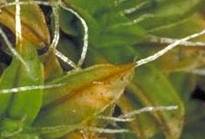
Moss and leafy liverwort leaves are generally just one cell thick. A nerve is an area of the leaf that is more than one cell thick and so often shows quite clearly, sometimes even to the naked eye if you hold a leaf up to the light. Whenever you're checking for a nerve make sure you are looking at a moist, open leaf. Nerves come in quite a variety of forms and the following stylized diagrams show you some, but not all, of the variety. In each case the nerve is shown in black.
The leftmost diagram depicts a situation similar to that shown in the photograph above – a narrow nerve that runs the length of the leaf's axis and extends beyond the leaf. In the next diagram the nerve extends to the leaf apex, but not beyond. Then follow two examples of successively shorter nerves and the fifth diagram shows a leaf with a short, forked nerve. The final diagram shows a leaf with a very wide nerve. The following drawing, kindly provided by Judith Curnow, shows a cross-section of a leaf of the moss in the genus Barbula. You can see that the leaf is largely one cell thick, but there is a clear thickening (the nerve) in the centre. The nerveless part of the leaf is about a hundredth of a millimetre thick.
While moss leaves are, apart from a nerve, generally one cell thick there are some exceptions, such as the genus Leucobryum. The following diagram shows a stylized depiction of a transverse cross-section of a Leucobryum leaf - thick in the middle and narrowing towards the margins. There are two rows of colourless, empty cells, a row of small, chlorophyllous cells (marked red) in the middle - but no nerve.
The preponderance of colourless cells gives the species of Leucobryum a relatively pale appearance - from pale green to whitish. Here ![]() is a photo of a colony of Leucobryum subchlorophyllosum growing on a fragment of tree fern stem on Norfolk Island. Here
is a photo of a colony of Leucobryum subchlorophyllosum growing on a fragment of tree fern stem on Norfolk Island. Here ![]() is a closer view of the same colony and here
is a closer view of the same colony and here ![]() , yet closer, is a different Leucobryum.
, yet closer, is a different Leucobryum.
In the leaves of some leafy liverworts you will find a feature called a vitta. This is a strip along the leaf's central axis, composed of enlarged cells. The vitta is still just one cell thick, so it is quite distinct from a nerve in a moss leaf which is more than one cell thick. Where the leaf is just a single cell thick the cells will show clearly, at the appropriate magnification. Because a nerve is two or more cells thick it can be hard to see all the overlapping nerve cells clearly, whereas there is no such difficulty in the case of a vitta . In some cases you can spot the difference between the nerve and non-nerve areas with a hand lens, but there are times when you would need a microscope to be sure. Also take care not to mistake the thalli of some of the simple thallose liverworts for moss leaves. For example, if you look at this photo ![]() of the simple thallose liverwort Symphyogyna podophylla you might think this is a moss in which the leaves have nerves. In fact, if you looked at the whole gametophyte carefully you'd see that there is no leaves-on-stem growth form. There is a thickened midrib, but it is a midrib through a thallose gametophyte.
of the simple thallose liverwort Symphyogyna podophylla you might think this is a moss in which the leaves have nerves. In fact, if you looked at the whole gametophyte carefully you'd see that there is no leaves-on-stem growth form. There is a thickened midrib, but it is a midrib through a thallose gametophyte.
The earlier photo (and the first of the diagrams) showed cases where the nerve in the moss leaf extended as a white hairpoint beyond the leaf apex. Just because you see a leaf with a hairpoint don't assume that a nerve is present. It may be that the leaf itself tapers to a long, hair-like apex and there is no protruding nerve. Always check the body of the leaf for the presence of a nerve.
If you see a definite nerve with the naked eye or with the help of a hand lens, well and good. If you don't it may be that there is no nerve or there is nerve, but one that is very weak or very short and so hard to see without the help of a microscope. It can sometimes also be hard to realise a nerve is present if you have species with a very wide nerve. In some cases the nerve may occupy over two thirds the leaf width and, if you're inexperienced, it can be easy to overlook the narrow "wings" on either side of the nerve. However it pays to put effort into checking for a nerve because the presence of a nerve carries a lot of information: If a nerve is present the bryophyte is definitely a moss.
If there is no nerve, the bryophyte in question can be a moss or a liverwort and you then need to look for other features to determine which bryophyte you have.
The rest of this page is devoted to some diagrams illustrating some of these leaf features. The moss leaves will be illustrated in black-and-white, the illustrations taken from Analytical Drawings of Australian Mosses, edited by Ferdinand von Mueller and published in Melbourne in 1864. The coloured illustrations of liverwort leaves have been taken from Die Lebermoose Deutschlands, by Gotthold Hahn, published in Germany in 1885.
If the leaf is notched, bears finger-like projections or is heavily divided the bryophyte in question is a liverwort. Otherwise it may be either liverwort or moss. You can often see such leaf shapes with a hand lens but there certainly are cases where you'd need to use a microscope to be sure.
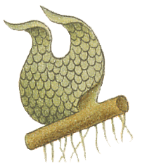 Jungermannia curvifolia |
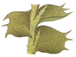 Jungermannia quinquedentata |
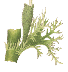 Trichocolea tomentella |
When leaves are heavily divided they give the whole plant a somewhat fuzzy appearance as shown by these photos of the leafy liverworts Chaetophyllopsis whiteleggei (close up ![]() and a little drawn back
and a little drawn back ![]() )and Leiomitra lanata (a fairly close view
)and Leiomitra lanata (a fairly close view ![]() and drawn back
and drawn back ![]() ).
).
 Note that in some moss species the leaves have very short teeth or slight serrations (as shown in the illustration of a Dawsonia leaf, right) or even short hairs. You can see another example of a moss with toothed leaf margins on the RACOMITRIUM PRUINOSUM page. In mosses the leaf shape varies from lanceolate to almost circular, depending on the species.
The cells of leafy liverworts are typically isodiametric. In moss leaves there may be areas with isodiametric cells, but elongated cells typically dominate. In the following pair of illustrations, on the left are several leaves of Calypogeia trichomanis and on the right is part of a leaf of Tortula antarctica. In that moss leaf, the dark band towards the left hand edge is the costa and on the right the leaf margin is folded in a little.
 Calypogeia trichomanis |
 Tortula antarctica |
Leafy liverworts typically have two distinctly different types of leaves. There's typically two rows of larger leaves, the lateral leaves, that extend out from each side of the stem and a third row, called underleaves, running along the stem. The underleaves are typically much smaller than and usually of quite a different shape to the lateral leaves. Such lateral and under leaves of markedly different forms are very rare in mosses. If you look at the left hand illustration in the previous pair you'll see two very small forked underleaves as well as three lateral leaves.
Liverwort leaves contain oil bodies ![]() , which are distinct, membrane-bound cell organelles with oily content, quite distinct from the green, chlorophyllous chloroplasts. Moss leaves lack such oil bodies. You need a microscope to see the oil bodies and they are best searched for in fresh specimens since the oil bodies may break down in specimens that have been collected and dried for long-term preservation in herbaria.
, which are distinct, membrane-bound cell organelles with oily content, quite distinct from the green, chlorophyllous chloroplasts. Moss leaves lack such oil bodies. You need a microscope to see the oil bodies and they are best searched for in fresh specimens since the oil bodies may break down in specimens that have been collected and dried for long-term preservation in herbaria.
Near the two basal corners of a moss leaf are the alar cells. These cells are typically thin-walled and larger than cells elsewhere in the leaf. Alar cells control the leaf's movement. When the cells are hydrated and swollen they tend to force the leaf away from the stem. When the alar cells lose water in dry periods they collapse, which tends to pull the leaf closer to the stem. The alar regions vary in size and number of cells between species and there is also variation in the extent of leaf movement between species.
This photograph ![]() shows part of a leaf of the moss Wijkia extenuata. The ragged edge on the left shows where the leaf had been attached to the stem. That raggedness arose when the leaf was pulled away from its stem. You can see several alar cells in the upper left quarter of this photo. When the leaf was mounted for photography part of it folded over itself so that the other alar region of this leaf is also in the photo, a little to the left below the centre of the photo. It is certainly not as clearly visible as the one to the upper left. You may also see the other leaf edge as a curved, somewhat thicker line, running to the lower right corner of the photo.
shows part of a leaf of the moss Wijkia extenuata. The ragged edge on the left shows where the leaf had been attached to the stem. That raggedness arose when the leaf was pulled away from its stem. You can see several alar cells in the upper left quarter of this photo. When the leaf was mounted for photography part of it folded over itself so that the other alar region of this leaf is also in the photo, a little to the left below the centre of the photo. It is certainly not as clearly visible as the one to the upper left. You may also see the other leaf edge as a curved, somewhat thicker line, running to the lower right corner of the photo.