Two Groups - classifying fungi into ascomycetes and basidiomycetes:
Mushrooms
Mushrooms are the best known basidiomycetes. Let's start with a mushroom and look at it's structure, at three different levels of magnification. Suppose you slice vertically across the outer part of a mushroom cap and pull away the small off-cut. The following diagram shows what you'd see.
 |
 Section through Hypholoma arantiacum cap |
For interest's sake, the diagram represents a mushroom with concentric rings of short, black scales on the cap. In most mushrooms the gills are V-shaped in cross-section, with the point of the V at the bottom edge of the gill. The grey colour represents the exposed flesh of the cap and gills. In some cases you'd need to use a hand lens, that magnifies 10 to 20 times, to clearly see the V shape of the gill cross-section.
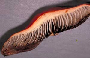
The V-shape of the gill cross section is exaggerated in the diagrams. The photo shows a slice taken from the cap of a mushroom of Hypholoma aurantiaca. You can see the white flesh that makes up the relatively narrow V-shape in each of the gill cross-sections. The gill faces have numerous basidia bearing dark brown spores - hence the dark coloured areas on either side of each gill section.
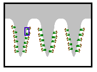
The gills of a mushroom are lined with basidia. So, if you took a very thin slice, across several gills, from that exposed section, and viewed it at about a hundred times magnification you'd see the basidia sticking out from the gills, as shown below - though in most mushrooms the top of the V would be much narrower than shown here. The basidia are drawn in green, the spores in brown and the general gill tissue again in grey. These colours (and those in later diagrams) have no significance and are simply used to differentiate the various structures. The basidia protrude from one gill into the airspace between that gill and the neighbouring gill. During their development, the spores are exposed to the air between the gills - quite unlike the ascomycetes, where the spores are enclosed in asci.

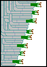
If you took a small section from a gill (such as the area contained in the blue rectangle in the above figure) and looked at it under the microscope at a magnification of several hundred times, you'd see something resembling the next figure. You can now see the basidia much more clearly and also see the hyphae that make up the gill tissue. In this diagram the hyphae are marked by the parallel pairs of greyish-blue lines and you can also see the septa that divide the filamentous hyphae into separate compartments. Notice that the basidia are the terminal elements of various hyphae and that between the basidia there are numerous ordinary hyphae. You can follow the hyphae back from the basidia and see that they bend upwards and may weave under and over each other.

There is much variation in shape of basidia and arrangement of hyphae in the gills.
![An Australian Government Initiative [logo]](/images/austgovt_brown_90px.gif)

